
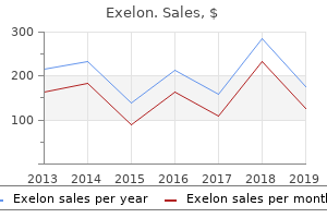
Exelon
Discount 3mg exelon fast delivery
Most ependymomas may be rather well circumscribed and may present a relatively clear surgical plane for resection symptoms 7 days after embryo transfer generic 6 mg exelon amex. Neuromonitoring with somatosensory evoked and motor evoked potentials may also be utilized depending on the surgical indication or surgeon preference symptoms heart attack discount 3 mg exelon mastercard. Although the vertebral level of the sternal angle varies between T2 and T7, it is most commonly at the T4 or T4-T5 intervertebral disk. If reduction cannot be achieved, a 540-degree procedure may be necessary in some cases (depending on the pathology), whereby the posterior approach and incision is temporarily closed and the patient is moved to a supine position for a ventral decompression followed by reopening of the posterior incision and posterior fixation. The use of intraoperative electrophysiology for the placement of spinal cord stimulator paddle leads under general anesthesia. The sternohyoid and sternothyroid muscles are divided near their sternal insertions. Coaxial double-lumen methylmethacrylate reconstruction in the anterior cervical and upper thoracic spine after tumor resection. Although navigation systems appear accurate, fundamental knowledge of navigation principles is needed to perform this technique adequately and safely. The dorsal and ventral nerve roots may already be contained within a common arachnoid sheath at the proximal origin of cauda equina tumors. Granulation tissue known as rheumatoid pannus then forms in the affected joint as a result of proliferating fibroblasts and inflammatory cells. As most of the lateral transthoracic approaches to the spine involve some type of fusion, the rib can be saved to be used as a bone graft. However, in recent decades, advanced surgical techniques and new approaches have led to a significant decrease in the associated surgical morbidity and mortality. The exam is usually normal even when intermittent neurologic symptoms are reported. Patients subsequently experience decreased pain and require shorter hospital stays. Frequently, successful posterior fusion and instrumentation at this level posteriorly leads to eventual arthrodesis anteriorly as well. This approach enables a C1 anterior arch arthrodesis to C3 using structural support graft and instrumental screw/plate fixation for a one-stage stabilization following odontoidectomy. Connectors used between segments of the shunt should be secured with a ligature placed around the tubing over the groove in the connector. The deformity is characterized by a lateral tilt of the head with rotation of the face. Often these tumors are quite sizable at the time of diagnosis, with little or no objective neurologic deficit. A thorough history should also address previous trauma, athletics, and activity involvement. In relatively simple cases, such as endoscopic sympathectomies, only two ports are required: one port for the endoscope and one port for the working instrument. Computed tomography-guided percutaneous cordotomy for intractable pain in malignancy. During the anterolateral dissection of the vertebral body, a wrapped sponge in the form of a cylinder is used to progressively separate the great vessels from the spine. The use of locally harvested bone from the lamina and medial facets provides autograft without the morbidity of harvest at a distant site, but may be inferior to uniform cancellous marrow from the iliac crest. They may present as incidental findings on imaging studies or present with profound neurologic deficits. The disease cannot be characterized by its presentation pattern, and the therapeutic options range from conservative to surgical.
Generic exelon 6 mg overnight delivery
Maximal benefit can be realized when patients are treated early in the disease process medicine 752 best order exelon. Additionally medications for high blood pressure order exelon line, the first, 11th, and 12th rib heads articulate only with their own vertebral body. Interdental fixation is required at the end of the procedure; therefore, an oral endotracheal tube is not an option. Biomechanical study of screws in the lateral masses: variables affecting pull-out resistance. After the surgical indication has been addressed, the posterior nasopharyngeal incision is not reapproximated. The endotracheal tube is secured in place and three-point head fixation is applied. Bipolar electrocautery and Gelfoam (Pharmacia & Upjohn Company, division of Pfizer Inc. The second most commonly seen anomaly is malrotation of a normally functioning kidney. The lateral wall is resected in a wedge shape using a Leksell rongeur working toward the anterior aspect of the spine. Intraoperative microscopy can make thecal compression safer and more precise and should be considered based on the pathology. Pain Cancer Pain More than half of the patients with pain due to malignancy may be undertreated. Traditionally, cervical stenosis and laterally herniated disks were treated through standard open procedures that consisted of posterior laminoforaminotomies with or without diskectomy. Proper positioning of the face requires collaboration between the surgeon and anesthesiologist, to make sure no additional pressure is placed on the eyes. It involves resection of the rib and transverse process to provide greater exposure to the ventral aspect of the thecal sac than other posterior approaches to disk pathology. Following skin incision, a Kelly clamp is used for dissection through the anatomic plane of the external and internal obliques and through the. Angiography is useful in delineating spinal vascular anatomy-such Symptoms Thoracic spine tumors present in a variety of ways based on their location within the vertebra and the speed with which they grow. The ligamentum flavum can then be identified and removed to expose the dorsal epidural pathology. Two-hole plates are ideal for patients with single-level subluxations or facet dislocations. These tumors are most commonly found outside the central nervous system, and when they occur in the spine (uncommonly) they tend to be extradural tumors. The radicular artery is located in this region, and care is taken not to damage it. On the other hand, the endoscopic transcervical approach provides sterile surgical corridors through the neck to the entire cervical spine via a beveled-tip tubular retractor. These lesions are more likely to be associated with hematomyelia or subarachnoid hemorrhage, which may be evacuated intraoperatively. Multiple systems are commercially available, and they have great appeal to surgeons unwilling to accept radiation exposure. Although the Wiltse approach has the advantage of sparing the posterior spinal elements from destabilization, it does not allow for central decompression (which is rarely needed). Patient Selection, Indications, and Contraindications Percutaneous methods are applicable to patients with a variety of pathologies. Technique the patient is placed in the lateral decubitus position, with as much flexion as possible without obstructing access to the abdomen (Video 111. The wound is then irrigated with sterile antibiotic solution and closed in layers with absorbable suture. Although a neck mass may be palpable, this is not a sensitive physical examination finding and is definitely not the norm for tumors based in the vertebral body. For patients with < 50% anterolisthesis and a nonkyphotic lumbosacral angle, a single-level fusion is typically adequate. Pain control via a combination of opiates and nonsteroidal anti-inflammatory drugs can allow the patient to achieve a state of adequate ventilation. Rod fracture is not uncommon following three-column osteotomies and can be seen in up to 22.
Order exelon cheap online
These two features are often interpreted with caution in the grading of ependymomas medications education plans order 4.5 mg exelon overnight delivery. Soft tissue structures resume normal anatomic alignment when the retraction is removed medicine engineering buy line exelon. Frempong-Boadu the intervertebral foramen is bordered superiorly and inferiorly by two adjacent pedicles, anteriorly by the vertebral body and disk, and posteriorly by the pars interarticularis and superior articular process of the facet joint. Indeed, some individuals have an extremely large L5-S1 interlaminar space, making bone removal either unnecessary or minimal. Over time, nucleus pressure is reduced while compressive load-bearing by the annulus is increased. Once decompression is complete, a subfascial vacuum drain may be used, which is brought out through a rostral exit wound, especially in cases involving multiple levels. Frempong-Boadu A variety of disk herniations can be treated with a tubular re tractor approach (primary or recurrent, small or large, paracen tral or foraminal). Graft Selection In addition to meticulous preparation of the fusion surface, careful consideration should be given to the selection of graft material, as the biological properties of the graft affect the eventual development of a solid fusion. If possible, the epidural fat should be preserved, although there is often a paucity of fat in patients with very severe stenosis. There is some consensus in the literature that up to three contiguous segmental vessels can be ligated at one side with minimal risk of neurologic deficit due to vascular compromise of the spinal cord. Grading of muscle strength in the muscle groups innervated by the suspected affected nerves should be performed, and sensation including light touch, pin prick, and proprioception should be assessed. Obstruction at the level of the gastric outlet by a gallstone is called Bouveret syndrome. Cottonoids soaked with oxymetazoline are placed in each nasal cavity for decongestion. The posterior approach enables lamina decompression along with pedicle screw fixation. In this manner, "construct-to-construct" compression may be performed and this may reduce the likelihood of fixation failure. Minimally Invasive Thoracic Microendoscopic Diskectomy: Lateral Transforaminal Approach this technique utilizes tubular muscular dilators/retractors via a posterolateral approach along with drilling of the lateral facet complex with or without resection of the pedicle. We approximate the skin as in any other case (running absorbable subcuticular suture and skin adhesive). In what has become the most common classification system, Wiltse and Newman characterized spondylolisthesis based on presumed etiology into five main groups: congenital, isthmic, degenerative, traumatic, and pathological. Then in a suprafascial plane and laterally, the site for the dilators and retractor is opened. Care should be taken to ensure that the aorta and vena cava, which run just anterior to the vertebral body, are not injured while placing and opening the retractor. In the interest of minimizing radiation exposure to the surgeon and staff, utilization of navigation during the insertion phase of the procedure has been described. Unless these fascicles arise from critical cervical or lumbosacral levels and demonstrate intraoperative stimulation, they need not be preserved, as such futile dissection unnecessarily prolongs the resection. Some authors open the fascia sharply or with monopolar electrocautery, but in most circumstances the retractors adequately split the tissue. Whenever sympathetic lesions are being created, it is important to remember that the majority of the sympathetic tone provided to the heart is contributed by the left sympathetic chain. Preoperative selective intercostal angiography in patients undergoing thoracoabdominal aneurysm repair. In a midline myelotomy, look for angulated vessels passing into the midline raphe. C1-C2 transarticular screw fixation is a very useful technique that can be successfully employed by most spine surgeons. Initial Assessment and Stabilization Management of patients with suspected cervical spine injuries begins at the scene of injury with strict immobilization of the neck during extrication from the scene and transport to the hospital. Pelvic renal ectopia is usually unilateral and involves a normally functioning kidney.
| Comparative prices of Exelon | ||
| # | Retailer | Average price |
| 1 | Dollar Tree | 602 |
| 2 | Foot Locker | 569 |
| 3 | CVS Caremark | 230 |
| 4 | Starbucks | 958 |
| 5 | Hy-Vee | 144 |
| 6 | H-E-B | 133 |
Purchase exelon
Use of a hemostatic matrix down a heavily bleeding pedicle tract should be avoided symptoms kennel cough exelon 1.5 mg sale, as it has been associated with fatal embolism medicine 968 cheap exelon 1.5mg with visa. Arthrodesis is then performed using pieces of cancellous and corticocancellous iliac crest autograft, which may be secured with the ends of the wire. Due to the role of the muscles below C6 in shoulder girdle and upper limb movement, U-sutures should be used if repairing the musculature of the lower cervical spine, to allow for additional tensile strength. Each landmark is dissected free of its fascial sheath and preserved both anatomically and functionally. Placement of hardware in an infected surgical bed had been strongly discouraged in the past, and a small number of surgeons still defer to a staged procedure. Hemangiopericytoma invading the craniovertebral junction: First reported case and review of the literature. For example, if there is a subluxation at C4-C5 with an associated fracture of the lateral mass of C4, two-level fixation between C4 and C5 will immobilize this motion segment unilaterally only on the side of the intact lateral masses. Occasionally, the placode is too wide, and repairing the neural tube defect compromises the perfusion of the tissue. Bipolar cautery may be used to address discrete bleeding vessels, although it should be done only after ensuring separation from nearby thecal sac and nerve roots. If a nerve is found to be running directly over Closure Hemostasis is achieved and the retractor system is removed. Cauda equina syndrome after in situ arthrodesis for severe spondylolisthesis at the lumbosacral junction. Excessively large craniectomies have been reported to cause cerebellar sagging postoperatively. Pyogenic vertebral osteomyelitis: a systematic review of clinical characteristics. Cervical spondylosis includes all the degenerative changes associated with aging that affect the vertebral bodies, intervertebral disks, facet joints, and spinal ligaments. In general, the T1 pedicles tend to be larger with a more rounded cross section compared with the ovoid pedicles elsewhere in the thoracic spine. When rotation is present, special attention must be given to the dural opening to prevent injury to the underlying dorsal roots or placode. Division of the diaphragmatic crus from the anterior longitudinal ligament and subsequently of the arcuate ligament from the transverse process of L1 facilitates exposure of the caudal extent of T12. Port Placement Incisions for the ports are placed in either the anterior axillary or posterior axillary line. Classic night pain may also occur with these tumors, but this symptom may also occur with extradural primary or metastatic tumors of the spine. Extreme care must be taken when removing the lamina over the septum in this type of malformation because the traversing Dural Opening and Septum Resection of the Split Cord Malformation Type I (Diastematomyelia, Bony Spur) the midline bony septum, which is always located extradurally, should be left in situ after circumferential removal of the overlying lamina is performed. Often, the surgeon may encounter some epidural bleeding after adequately decompressing the nerve root. Additionally, a physical exam should be performed and imaging should be obtained to determine the functionality of the phrenic nerve. Purulent drainage is present on the buttocks from the sinus located at the inferior aspect of the subcutaneous area of redness and swelling. Additionally, neuronavigation systems may facilitate increased accuracy over free-hand and fluoroscopyassisted screw placement, with reduced surgeon exposure to radiation. Safety of spinal angiography: complication rate analysis in 302 diagnostic angiograms. All remaining soft tissues overlying the vertebral body should be cleared to identify the lateral borders of the decompression to ensure a symmetric, rectangular decompression. Because many patients are significantly compromised medically and neurologically, careful attention to nutritional and respiratory issues prior to surgery will help ameliorate problems after surgery. It should be noted that dissection of the right side can be undertaken safely if the right-sided approach is indicated. The absence of brain tissue within the sac is the single most favorable prognostic factor for survival. The tubular retractor is then secured to a table-mounted arm with downward pressure applied to keep muscle out of the field.
Discount exelon online amex
Other pathological conditions such as proatlas segmentation abnormalities medicine 877 purchase exelon 1.5 mg otc, atlantoaxial tumors medicine urology buy 4.5mg exelon with amex, clival tumors, and rare congenital osseous abnormalities can also result in ventral cervicomedullary compression. The patient underwent a partial T6 laminectomy and partial laminectomy of T5 and T7 for decompression. A complete resection of the lesion was achieved, sparing the rest of the fascicles of the posterior cord. Any maneuver that attempts to lengthen the spine puts the neural elements at significant risk. Use lateral fluoroscopy confirmation and guidance throughout the surgery; navigation is also an option. With this technique, the tulip head of the iliac screw is in line with lumbosacral rod placement. Preoperative spinal artery localization and its relationship to postoperative neurologic complications. Spontaneous regression of a thoracic calcified disc herniation in a young female: a case report and literature review. At this point a neuromonitoring probe is slowly passed through the psoas muscle under direct fluoroscopic guidance to dock at the intervertebral disk within one of the previously mentioned safe zones. The arch bars are used to place the teeth in interdental fixation at the end of the procedure. Except in cases of diskectomy without interbody fusion, we mobilize our patients with a thoracolumbar orthosis. Complications in patients with palmar hyperhidrosis treated with transthoracic endoscopic sympathectomy. In some cases an access surgeon, either a general surgeon of a vascular surgeon, may be needed to assist with the exposure, which requires mobilization of the great vessels and handling of the abdominal contents. Prospective, randomized, multicenter study of cervical arthroplasty: 269 patients from the Kineflex C artificial disc investigational device exemption study with a minimum 2-year follow-up: clinical article. After corpectomy has been completed (a), a titanium strut filled with autologous bone graft is seated firmly in the corpectomy site under distraction of the vertebral bodies above and below (b). The transversalis fascia and aponeurosis of the transverses abdominus and oblique muscles are repaired to prevent hernia formation. The ipsilateral transverse processes can be palpated in the crevice that is formed from the medial border of the quadratus lumborum with the lateral boarder of the psoas muscle. The patient underwent an urgent decompression and fusion due to his progressive neurologic decline. Collateral supply must be ruled out at the time of treatment by injections at the correspondent levels on the contralateral side, as well as adjacent segmental arteries above and below the fistula. In the pediatric patient, these conditions occur most commonly at the lumbosacral junction, involving the L5 pars followed by L4. The paraxial mesoderm forms condensations (somites) flanking the neural tube bilaterally at approximately day 25 of gestation. Ascaris lumbricoides and Opisthorchis sinensis are seen in patients with recurrent pyogenic cholangitis. The system provides a rational approach to surgical planning while taking into account the limitations of en-bloc excisions created by preservation of the cord. Pelvic incidence: a fundamental pelvic parameter for three-dimensional regulation of spinal sagittal curves. Cervical spine radiographs should be taken in children who present with torticollis to rule out osseous etiologies. Once the ligamentum flavum has been removed, the epidural space is explored with the purpose of identifying the nerve root. Also, the dystonia that is frequently associated with spasticity has dynamic characteristics. There are few or no sequelae associated with sacrificing the sensory nerve containing the neurofibroma. Slight craniocaudal angulation away from the expandable cage is acceptable, but excessively divergent trajectories should be used with caution as this may interfere with the screw locking mechanism (depending on the particular system) and increase the risk of construct failure. For example, paired bilateral hemivertebrae can result in a "balanced" scoliosis as the curves cancel out. After closure of the incisions in two layers and ap plication of sterile dressings, an abdominal binder may be placed in order to increase the resistance to any fluid accumulation under the pump site or spinal incisions. Preoperative assessment, including anesthesia considerations, helps minimize the risk of intraoperative complications.
Order exelon 3mg free shipping
If this is unsuccessful strong consideration should be given to C2 root transection medications diabetes cheap exelon american express. Processed allografts and type I collagen conduits for repair of peripheral nerve gaps 5 medications exelon 3 mg. This can be done later but should be performed before removing a substantial part of the vertebral body. Due to their expansile nature, they may cause swelling, pain, bone destruction, and fractures. It must be used carefully, though, due to the risk of congestion of irrigation flow and mismatching inflow and outflow. The lung is then reinflated, which ensures adequate reexpansion and ventilation of the lung. The actual technique for trial lead implantation does not differ from permanent lead placement (described below). A small dural defect can usually be identified as the cause of the persistent leak and can be primarily repaired with suture and a small muscle graft. For large defects, a closed suction drainage sys tem is often used to minimize the risk of postoperative hema toma. Urodynamic findings in the tethered spinal cord syndrome: does surgical release improve bladder function Occult spinal dysraphism: clinical and urodynamic outcome after division of the filum terminale. There are several reports of symptomatic patients with a normal-lying conus who improve after tethered cord release. When these lesions appear in the spine, they are typically located in the vertebral body but may expand to the surrounding cortical bone as the lesion grows. Vascularized rib may be used due to its accessibility, high content of bone morphogenic protein, and vascularity. In the recovery period, vomiting can be controlled by steroids or antiemetics, and the surgeon should have a low threshold to prescribe antiemetics to stay ahead of vomiting episodes postoperatively. A reverse Trende lenburg position or ankle weights may be used to counteract the pull from the traction weights. To control the springiness of the elevated lamina, the open side of the "open door" is drilled first. Compared with the traditional anterior approach, the retroperitoneal approach poses less risk to the peritoneal organs and great vessels. Rostral margin of the placode (white arrow) is continuous with the normal spinal cord. The presence of a large herniated disk fragment anterior to the cord would usually be handled with an anterior approach. Postoperative Care Postoperative care is fairly routine in patients undergoing the standard open retroperitoneal approach. Tonsillar herniation: the rule rather than the exception after lumboperitoneal shunting in the pediatric population. The trajectory is along the angle of the ipsilateral lamina, aiming for the intersection of the ipsilateral transverse process and superior facet. The anterior approach to the lumbosacral junction thus remains an essential and powerful tool not only for exposing the anterior and middle columns of the lumbosacral junction, but also for releasing rigid spinal deformities and achieving optimal lordosis restoration in the lumbar spine. The patient is secured to the table, and the spinal level is marked on the skin using anterior and lateral fluoroscopy. Surgical rates and operative outcome analysis in thoracolumbar and lumbar major adult scoliosis: application of the new adult deformity classification. Superiorly a subperiosteal exposure of sacral margins and coccyx from rostral to caudal helps prevent inadvertent rectal injury. A second skin incision should then be marked in a similar fashion on the contralateral side of the tension band at the caudal pole of the lesion. Surgical management of extreme lateral lumbar disc herniations: review of 138 cases. However, if C1 or C2 laminectomy is necessary or any of the previously discussed anatomic or alignment concerns are present, C1-C2 segmental fixation is chosen. Disks can either bulge (where the annulus is intact), herniate (where the annulus tears and the disk protrudes), or become sequestered (the herniation is completely separate from the disk).
Trusted 4.5mg exelon
Because the surgical field is relatively small medicine lake montana cheap exelon 6mg otc, illumination and magnification are needed in order to visualize the surgical field properly treatment menopause purchase generic exelon pills. In these cases the need of posterior fossa decompression can be eliminated by addressing the primary pathologies. Minimally invasive surgery treatment for thoracic spine tumor removal: a miniopen, lateral approach. Type I is an unstable fusion pattern, such as two long segments of block vertebrae with an intervening open disk space. It is our preference to use fluoroscopy when placing the screws, but some surgeons prefer to rely on navigation guidance, and a smaller subset use neither technique. The disk spaces above and below the vertebral body to be resected are identified and incised with a fine-tipped knife blade. The drill-tap should not be advanced further than 1 cm from the tip of the K-wire. It is even more important for the neurosurgical community not to lose the experience in performing these procedures in an era of device implants for neurostimulation and intrathecal drug delivery. The transversalis fascia is identified and the posterior portion is opened to access the retroperitoneal space. A dorsal occipitocervical fixation can then be performed in the reduced position with or without decompression. Meticulous removal of soft tissue and coagulation debris over the extent of the transverse process should be performed as well as decortication of the dorsal aspect of the transverse process. Decreased spasticity can reduce the development of contractures and joint deformities. With an experienced access surgeon, straightforward anterior exposures can be performed efficiently and with minimal blood loss and relatively low morbidity through a reasonably small incision. A thorough diskectomy at the rostral and caudal disk spaces enables identifying the end plates above and below the area of planned resection and helps to prevent inadvertent violation of the end plates during the corpectomy. Postoperative Care the standard postoperative care for spinal surgery is given, including monitoring urinary output, providing pain control, and starting physical therapy. The continuous advancements in our knowledge of those lesions, however, also translated into a rapid proliferation of several different classification systems. In terms of bony anatomy, ventrally, the cervical vertebra is composed of a vertebral body connected to a posterior neural arch that together enclose the vertebral foramen, which includes the spinal cord. To place bilateral translaminar screws, the starting points have to be slightly different cephalad-caudal, and the lateral trajectories must be slightly divergent, to avoid collision of the screws. Caudally, the surgical field is limited by the aortic arch and its branches at the T3 and T4 vertebrae. The segmental vessels are sacrificed proximally along their course, ensuring that there is a sufficient proximal vessel to hold the ligature without slipping off. The skin entry point is adjusted cranially or caudally to be precisely in line with the disk space. Several proposed models to conceptualize injury and assess stability have contributed in some way to the current understanding of lumbar trauma. An index finger of the opposite hand is placed intraorally in the retromolar area to palpate completion of the osteotomy. In addition, five new disk herniations were detected in four patients during the follow-up period. In addition to maintaining the physical integrity of the roots, the vascular supply to these small structures must be maintained. Knowing the precise dimensions and configuration of bone tissue is useful for selecting the location and depths of the troughs. It is generally a result of erosion or fracture of the odontoid process and may result in myelopathy secondary to spinal cord compression. If mesenchymal elements from surrounding mesoderm proliferate while the connection between cutaneous ectoderm and neural ectoderm is obliterated too early, intradural anomalies such as intramedullary lipomas and lipomyelomeningoceles20 occur. Stabilization of the atlantoaxial complex via C-1 lateral mass and C-2 pedicle screw fixation in a multicenter clinical experience in 102 patients: modification of the Harms and Goel techniques.
Generic 1.5mg exelon otc
The specific spinal techniques that can be utilized after securing this exposure are discussed in the previous chapters in this section medications excessive sweating buy exelon 3mg. The presence of any of these symptoms aside from pain is often an indication for surgical intervention administering medications 8th edition order exelon 1.5mg line. Disruption of these nerves causes dysregulation of the internal vesicular sphincters, resulting in ejaculation of sperm into the bladder rather than out of the penis. This procedure has been refined from the traditional open techniques and offers excellent relief of symptoms with good long-term outcomes. Any paraspinal muscle obscuring the bony anatomy is resected using a pituitary rongeur and monopolar cautery with a long, bent tip. Potential disadvantages are that this procedure is very complex, and it requires significant experience. Individual fascicles can be grafted using a perineurial technique; alternatively, if the entire cross section of nerve has been damaged, an epineurial repair is performed. Eventually the skin sloughs away from the midline suture associated with fat necrosis and exudate along the incision. It is recognized the even the smallest incision, placed in the midline, may give rise to a small, usually segmental, sensory deficit. Second, the abdomen is decompressed partially (Wilson frame) or completely (Andrew table). The plan for surgical correction is either one of stabilization or one of correction depending on whether the patient has presented prior to the onset of major deformity. Neuromonitoring is required to safely traverse the psoas muscle, so muscle relaxants and inhalational anesthetic agents must be minimalized. This technique has several uses, including serving as a rescue and revision option and the ability to place cortical screws at the same level as existing pedicle screws, thus permitting adjacent-level fusion without the need for extensive reexposure or removal of preexisting hardware. Incidence of spinal column deformity after multilevel laminectomy in children and adults. Surgical Technique Anesthesia and Positioning Following induction, a nasogastric tube is placed for gastric decompression. Resegmentation of the Vertebral Column the distinct nature of each somite (or primary segment) as a developing unit changes. Crosslinks are either rod-to-rod or screw-to-screw, depending on the available rod surface. Prompt identification is necessary to aspirate the material, irrigate with antibiotic solution, and close with suction drains if an infection is found. The construct should include three normal levels above the pathology and two normal levels below the pathology, and the incision should provide adequate exposure. The subcutaneous tissues are then retracted using a selfretaining Weitlaner retractor. In cases of trauma, benign lesions, or for patients with malignant tumors who have a relatively long life expectancy (> 2 years), the goal of anterior reconstruction is to provide three-column support, to correct or maintain appropriate alignment, and to promote bony fusion. Filum ependymomas and cauda equina nerve sheath tumors occur with about equal frequency. It has been reported that radicular pain responds better to surgery than nonradiating axial thoracic pain. Electrical stimulation will determine if this is a motor fascicle and still functional or if fascicles enlarged by tumor provide motor function. The first of these techniques was the C1-C2 transarticular screw fixation, which was reported in 1979 by Magerl. The insertion point is 3 to 4 mm medial to the lateral border of the pars interarticularis at the junction of the caudal border of the transverse process. Our main goal as surgeons is to respect healthy and functional tissue as much as possible. Once the bone removal is complete, a small amount of bone wax applied with a Penfield instrument will easily control bleeding from the bone.